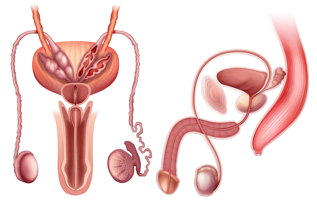
Structure and Anatomy of the Prostate

The prostate gland is a small, walnut-shaped gland that is located in the pelvis of men, just below the bladder and in front of the rectum. It is part of the male reproductive system and its main function is to secrete a fluid that nourishes and protects the sperm. The prostate gland surrounds the urethra, which is the tube that carries urine from the bladder out of the body.
The prostate gland consists of three main parts: the peripheral zone, central zone, and transition zone. The peripheral zone is the outermost layer of the prostate gland and is where most prostate cancers develop. The central zone is located in the middle of the prostate gland and is responsible for producing the majority of the prostate’s fluid. The transition zone is the area of the prostate gland that surrounds the urethra and is responsible for the enlargement of the prostate gland that occurs with age.
The prostate gland is made up of several different types of cells, including glandular cells, stromal cells, and smooth muscle cells. Glandular cells are responsible for producing the prostate’s fluid. Stromal cells provide support to the glandular cells and help maintain the structure of the prostate gland. Smooth muscle cells control the release of the prostate’s fluid during ejaculation.
The prostate gland is supplied with blood by several different arteries, including the inferior vesical artery, the middle rectal artery, and the internal pudendal artery. These arteries provide the prostate gland with the necessary oxygen and nutrients to maintain its proper function.
Understanding the structure and anatomy of the prostate gland is important for detecting and treating prostate cancer and other prostate-related conditions. Regular check-ups and screenings can help men maintain good prostate health and catch any potential problems early on.
