Anatomy of the Eye

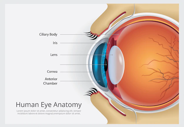
The eye is a complex sensory organ that allows us to see the world around us. It is composed of several structures that work together to capture and process visual information.
Cornea: The cornea is the transparent, dome-shaped outer layer of the eye that covers the iris, pupil, and anterior chamber. It plays a crucial role in focusing incoming light.
Iris: The iris is the colored part of the eye that surrounds the pupil. It controls the amount of light entering the eye by changing the size of the pupil.
Pupil: The pupil is the black circular opening in the center of the iris that allows light to enter the eye.
Lens: The lens is a clear, flexible structure that sits behind the pupil and helps to focus light onto the retina.
Retina: The retina is the light-sensitive layer of tissue that lines the back of the eye. It contains photoreceptor cells called rods and cones that convert light into electrical signals that are sent to the brain via the optic nerve.
Optic nerve: The optic nerve is a bundle of nerve fibers that carries visual information from the retina to the brain.
Vitreous humor: The vitreous humor is a gel-like substance that fills the center of the eye and helps to maintain its shape.
Sclera: The sclera is the white, outer layer of the eye that covers most of the eyeball. It provides support and protection for the inner structures of the eye.
Choroid: The choroid is a layer of blood vessels that lies between the retina and sclera. It supplies nutrients and oxygen to the retina.
Ciliary body: The ciliary body is a muscular structure that surrounds the lens and helps to control its shape, allowing the eye to focus on objects at different distances.
Overall, the anatomy of the eye is a highly specialized and complex structure that allows us to see the world around us in great detail. Each part of the eye plays a vital role in the process of vision, from capturing light to transmitting visual information to the brain.
